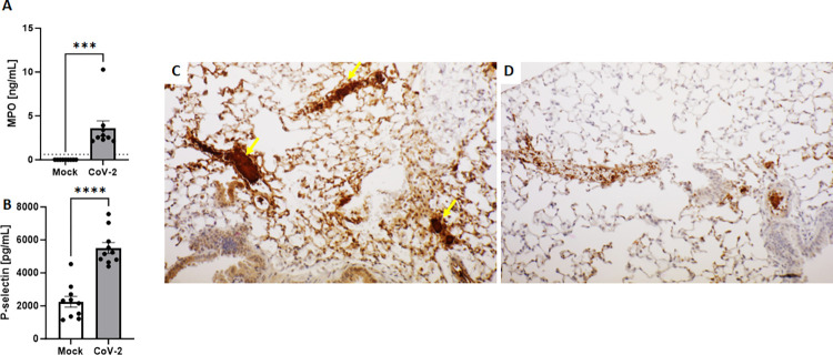
Fig 5. Lethal SARS-CoV-2 infection induced neutrophil/NETs and platelet/endothelium activation in AC70 hACE2 Tg mice.
Evaluation of neutrophil/NETs and platelet/endothelium activation (A&B). Quantification of myeloperoxidase (MPO) (A) and P-selectin (B). Gamma irradiated EDTA-plasma samples collected from Mock mice and 4 d.p.i. with 105 TCID50 of SARS-CoV-2 were analyzed by ELISA assays. Data are presented as means ± SEM (n = 5 per group). Dotted horizontal line indicate the limit of detection for MPO assay (0.61 ng/mL). Results were considered significant with p < 0.05 ***p = 0.0008, ****p<0.0001). Representative immunohistochemistry (IHC) staining of lungs to Platelet Factor-4 (PF4) (C&D). Activation of platelets is shown as brown staining. Completely blocked veins and capillaries in the alveolar walls of the infected lungs (C, yellow arrows) were seen compared to the non-infected tissues (D). Bar scale: 80μm. Combined data from two independent experiments.

