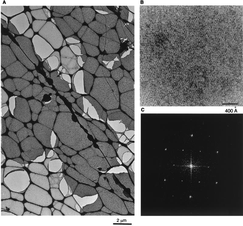
FIG. 6.
Assembly of His-MoCANC proteins on a lipid membrane. His-MoCANC proteins were incubated beneath PC-plus-Ni2+-DHGN monolayers overnight at 30°C, after which the membranes with bound proteins were lifted onto lacy EM grids, washed, and stained with uranyl acetate. (A) Stained protein arrays appear as homogenous dark-staining layers which cover the lacy carbon grid support (thick dark lines). Also visible are membrane areas devoid of protein (light grey) and lighter lacy holes indicative of broken membranes. Bar, 2 μm. (B) Shown is a high-magnification image of His-MoCANC proteins arrayed on a lipid monolayer, in which protein areas appear white. Bar, 400 Å. (C) The diffraction pattern of a His-MoCANC protein array was calculated as described in Materials and Methods and is displayed as a power spectrum. The pattern can be indexed in either hexagonal or orthorhombic fashion. The six innermost reflections correspond to the 1,0; 0,1; −1,1; −1,0; 0,−1 and 1,−1 reflections for a γ* = 60° unit cell and correspond to real-space unit cell edges of 77.2 Å.

