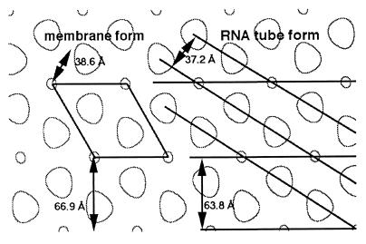
FIG. 8.
Comparison of His-MoCANC membrane-bound and RNA tube forms. Real-space His-MoCANC membrane-bound and RNA tube forms are compared on an edge-enhanced representation of a 2D crystal reconstruction, where small circles and larger off-circles indicate protein-free cage holes. On the left is shown a 2D crystal unit cell, along with the distances between the 1,0 (66.9 Å) and 1,1 (38.6 Å) planes (not corrected for γ = 120°). On the right are shown the basic helices of the RNA tube form, where a and b helices are separated by 63.8 and 37.2 Å, respectively. The tube form can be generated from the 2D net by rotating the 2D ab plane slightly and rolling the net into a tube.

