Figure 2.
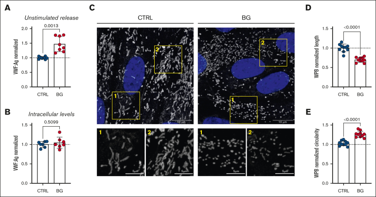
O-linked sialylation influences VWF secretion and alters WPB morphology. (A) Unstimulated VWF:Ag secretion levels from HUVECs incubated with or without BG (2 mM for 72 hours; n = 8 from 4 independent experiments; Welch t test; P = .0013). (B) VWF:Ag levels in unstimulated HUVEC cell lysates after treatment with or without BG (n = 8, from 4 independent experiments; t test; P = .5099). (C) Immunofluorescent images of HUVEC treated with BG (2 mM for 72 hours) compared with untreated CTRLs (VWF in gray; DAPI [4′,6-diamidino-2-phenylindole] in blue; representative images of 3 independent experiments); scale bars are set at 10 μm for overview images and at 5 μm for the zoomed regions; (D) Automated assessment of WPB length; results are depicted as normalized to CTRL (n = 10 images, from 3 independent experiments; t test; P < .0001). (E) Automated assessment of WPB circularity (data are shown as normalized to CTRL) in CTRL and BG-treated cells (n = 10 images, from 3 independent experiments; t test; P < .0001).

