Figure 5.
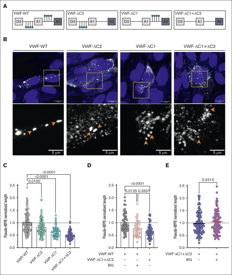
O-glycan clusters in VWF affect pseudo-WPB morphology. (A) Schematic representation illustrating OLG clustered around the VWF-A1 domain and the recombinant OLG mutants generated. (B) Immunofluorescent images of HEK293 cells expressing VWF-WT, VWF-ΔC2, VWF-ΔC1, and VWF-ΔC1+ΔC2. Orange arrowheads in zoomed images point to pseudo-WPBs (representative images of n = 3). Scale bars are set at 10 μm for overview images and at 5 μm for the zoomed regions. (C) Pseudo-WPB length in VWF-WT, VWF-ΔC2, VWF-ΔC1, and VWF-ΔC1+ΔC2 expressing HEK293 cells (n = 3; Kruskal-Wallis test; P = .01; P < .0001; and P < .0001). (D) Pseudo-WPB length in VWF-WT, VWF-WT treated with BG, and VWF-ΔC1+ΔC2 expressing HEK293 cells normalized to VWF-WT (n = 2 independent experiments; Kruskal-Wallis test with multiple comparisons; P = .0125; P < .0001; P = .3331). (E) Pseudo-WPB length in VWF-ΔC1+ΔC2 and VWF-ΔC1+ΔC2 treated with BG expressing HEK293 cells normalized to VWF-ΔC1+ΔC2 (n = 3 independent experiments; Mann-Whitney test; P = .0125; P < .0001; P = .3331).

