Figure 6.
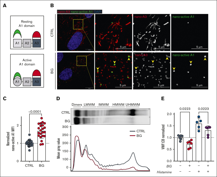
OLG inhibition leads to VWF-A1 domain activation and reduced HMWMs. (A) Figure illustrates use of nanobodies to detect activated VWF-A1 domain (nano-active A1 in green) vs normal VWF-A3 domain (nano-A3 in red). (B) Immunofluorescence images of nanobody interactions with BG-treated HUVECs compared with untreated CTRLs (VWF-A3 in red; active-A1 in green and DAPI in blue; representative images of n = 3). Yellow arrowheads point to WPBs positive for both inactive and active VWF. Scale bars are set at 10 μm for overview images and at 5 μm for the zoomed regions; (C) Mean fluorescence intensity (MFI) for nano-active A1 binding in BG-treated HUVECs vs untreated controls (n = 22, from 4 independent experiments; Welch t test; P < .0001). (D) VWF multimer blot and densitometry of conditioned media from BG-treated HUVECs compared with untreated CTRLs (representative images of 2 independent experiments). (E) VWF:CB for VWF secreted from HUVECs incubated with or without BG (1) under steady-state conditions and (2) after histamine stimulation (n = 6 from 3 independent experiments; 1-way analysis of variance [ANOVA] with multiple comparisons; CTRL vs BG, P = .0223; CTRL his vs BG his, P = .0223). his, histamine; IHWM, XXX; LHWM, XXX; UHWM, XXX.

