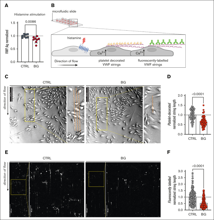
Figure 7.
Truncation of OLG causes reduces VWF strings on activated ECs. (A) VWF:Ag secretion after histamine (100 μM) stimulation from HUVECs incubated with or without BG (n = 8, from 4 independent experiments; t test; P = .0086). (B) Histamine activation of HUVECs under shear results in the production of tethered VWF strings on the EC surface, which can be detected using platelets or fluorescent anti-VWF antibodies. (C) Platelet-decorated VWF string visualized in brightfield after histamine stimulation of BG-treated HUVECs compared with untreated CTRLs (representative images of 2 independent experiments; direction of the flow from top to bottom; flow rate, 1.5 mL/min [2 dyn/cm2]; scale bars, 250 pixels). (D) Platelet-decorated VWF string length for HUVECs treated with BG compared with untreated CTRLs (Mann-Whitney test; P < .0001). (E) Fluorescently labeled VWF strings after histamine stimulation of BG-treated HUVEC compared with untreated CTRLs (representative images of 2 independent experiments; direction of the flow from top to bottom; flow rate, 1.5 mL/min [2 dyn/cm2]). (F) Fluorescently labeled VWF string length for HUVECs treated with BG compared with untreated CTRLs (Mann-Whitney test; P < .0001).

