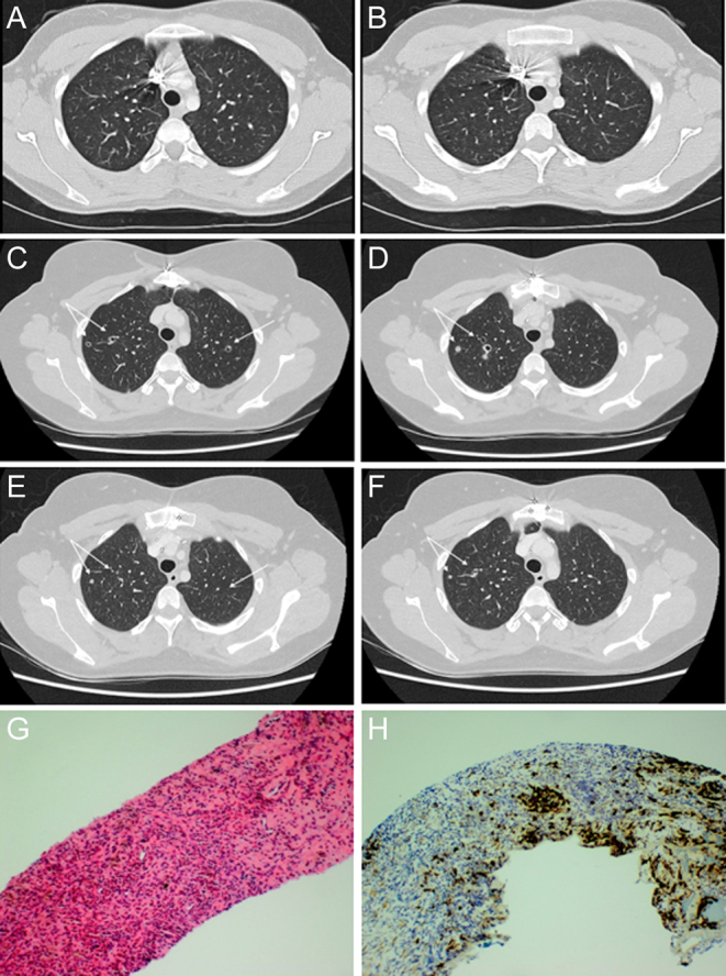
Figure 2.

Cavitating apical lung nodules pre- and post-inhaled corticosteroids and surgical pathology of the lung biopsy. Axial-view CT scan of the chest demonstrates bilateral cavitating subcentimetric apical nodules (C, D) which were not present on the initial CT scan pre-selpercatinib (A, B). There was almost complete resolution 3 months later with inhaled glucocorticoids (E, F). Serial haematoxylin and eosin (H&E) (G) and Langerin immunohistochemistry (IHC) (H) stained sections of the lung core biopsy demonstrating Langerhans cell histiocytosis (LCH). (G) On H&E, the Langerhans cells are visible but obscured by an infiltrate of chronic inflammatory cells rich in eosinophils. (H) Langerin IHC highlights the Langerhans cells which account for about 5% of the cells in the section (original magnification 100×).

 This work is licensed under a