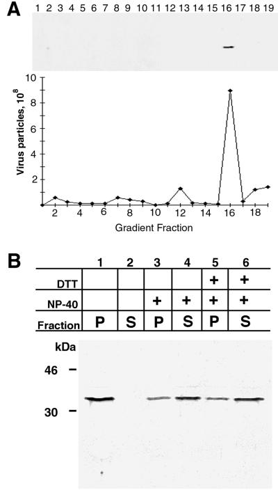
FIG. 4.
Association of the H3L protein with purified vaccinia virus particles and extraction with nonionic detergent. (A) Western blot of purified virus particles. Sucrose gradient-purified intracellular virions were centrifuged in a performed CsCl gradient. Fractions (0.5 ml) were collected from the top of the tube, diluted, and centrifuged to pellet virus particles. The pellets were suspended, and part of the suspension was used to calculate the number of virus particles from the absorbance at 260 nm; the remainder was analyzed by SDS-PAGE and immunoblotted with H3L peptide antibody followed by anti-rabbit IgG horseradish peroxidase conjugate. CsCl fraction numbers are indicated. (B) Extraction of H3L protein with NP-40 detergent. Purified vaccinia virions were incubated in Tris buffer containing 0.5% NP-40 or 0.5% NP-40 and 50 mM DTT. After centrifugation, the supernatant (S) and pellet (P) fractions were analyzed by SDS-PAGE and immunoblotting with the H3L peptide antibody. The masses and positions of markers are indicated at the left.

