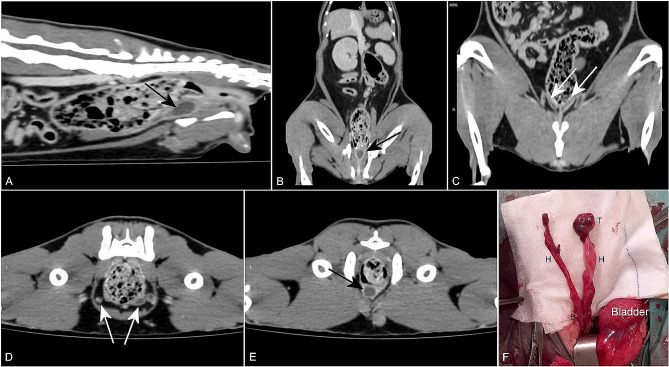
Fig. 2.
Postcontrast CT images (A, B, C, D and E) and the gross appearance (F) of the anomalous structures identified, adjacent to the descending colon. (A) and (B) A sagittal and a dorsal CT image, respectively, show a tubular structure with a high uptake wall, ventral to the descending colon (black arrow). (C) and (D) A dorsal and a transverse CT image, respectively, show the ramification into two branches (white arrows) of the previous identified structure. (E) A transverse CT image shows a nodular structure in the left inguinal subcutaneous tissue and a tubular structure, with a high uptake wall and a low uptake interior, ventral to the descending colon, compatible with a remnant uterus (black arrow). (F) Intraoperative image of the structures found. The two tubular structures (H) converge into a wider structure similar to a uterine body (B). At the end of one tubular structure, there is a small structure that resembles a testicle (T)

