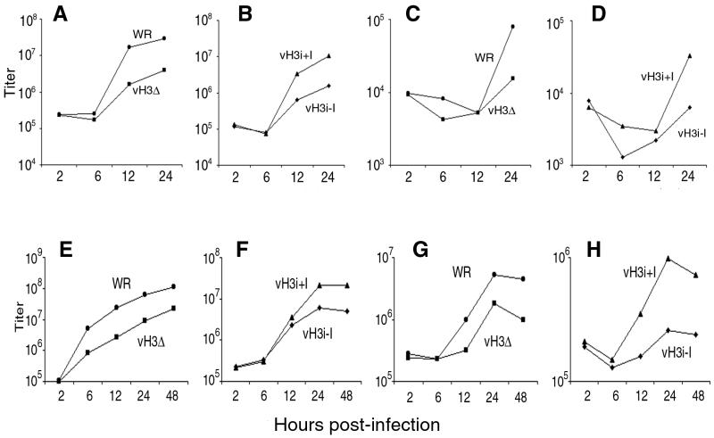
FIG. 3.
One-step growth curves. BS-C-1 cells (A to D) and RK13 cells (E to H) were infected with purified WR, vH3Δ, or vH3i in the presence (+I) or absence (−I) of IPTG as indicated. At 2, 6, 12, 24, and 48 h, the medium (C, D, G, and H) and cells (A, B, E, and F) were harvested separately and infectious virus titers were determined by plaque assay.

