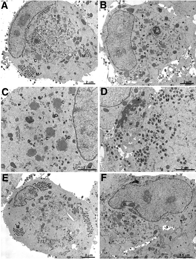
FIG. 6.
Electron microscopy of ultrathin sections of infected cells. RK13 cells were infected with WR (A), vH3Δ (B to D), or vH3i in the presence (E) or absence (F) of IPTG and incubated for 23 h. Abbreviations: C, crescents; I, IV; M, mature virions; D, DNA crystalloids. Magnification is indicated by bars.

