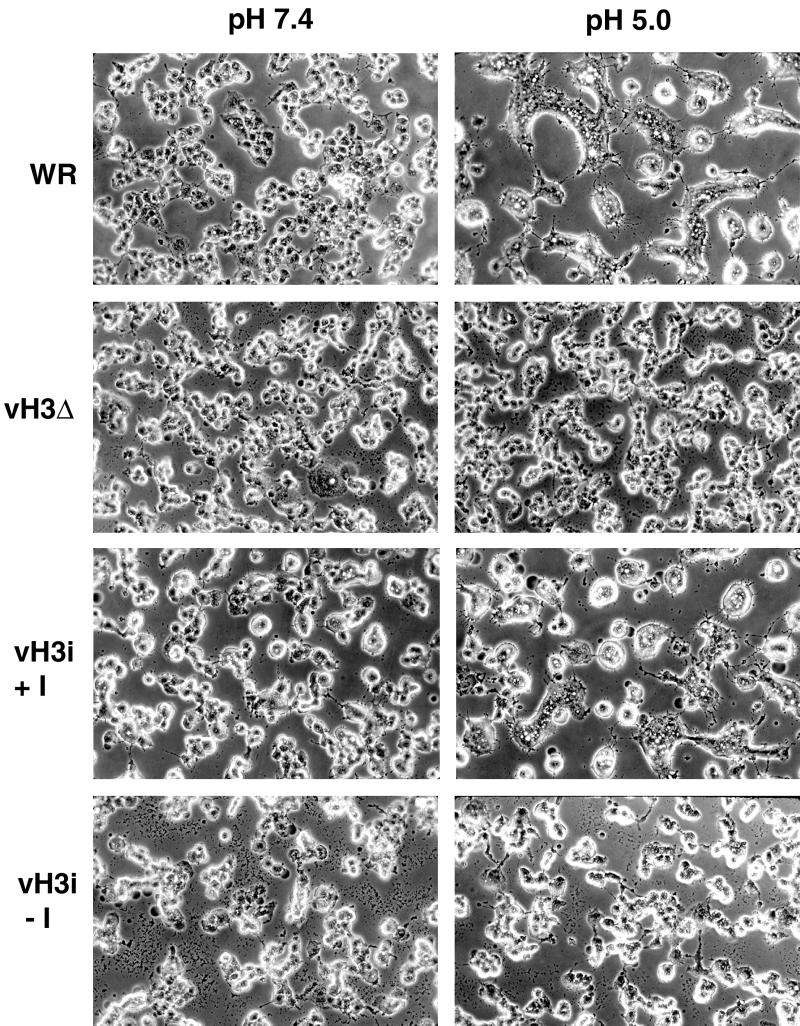
FIG. 9.
Low-pH-induced fusion of infected cells. BS-C-1 cells were infected with WR, vH3Δ, or vH3i in the presence (+I) or absence (−I) of IPTG. At 12 h after infection, the cells were immersed briefly in buffer at pH 5.0 or 7.4. The medium was replaced, and the incubation continued for an additional 3 h. The cells were photographed with a phase-contrast microscope.

