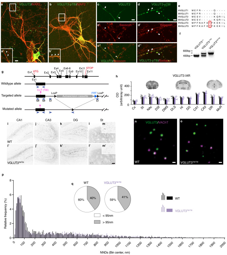
Fig. 2. The p.T8I variant does not alter the anatomical distribution of VGLUT3.
a-d, Immunofluorescent detection of VGLUT3 (a, a’, c, c’, c”) or VGLUT3-p.T8I (b, b’, d, d’, d”) (green) and microtubule-associated protein (MAP2, a, a’, b, b’) or bassoon (c’, c”, d’, d”) (red) in hippocampal neuronal culture of WT mice. e, Alignment of human (H) or mouse (M) VGLUT1, VGLUT2 and VGLUT3 amino acid sequences. f, Genotyping of WT (VGLUT3+/+) mice, heterozygous mice (VGLUT3+/T8I) or homozygous mice (VGLUT3T8I/T8I). g, Schematic representation of the targeting strategy and genotyping strategy of mouse VGLUT3 by PCR. Mice were genotyped with primers (arrows) flanking exon 1 (mf, mr, ex1), in intron 1 (ef) and in the LoxP sites in 3’ of the autoexcision cassette (er). Asterisks represent the targeted site in exon 1. h, Top: Immunoautoradiographic (IAR) regional distribution of VGLUT3 and VGLUT3-p.T8I on coronal sections from the brain of WT mice (black, n = 7 mice) and VGLUT3T8I/T8I mice (purple, n = 8 mice). Bottom: Densitometric quantification of VGLUT3 and VGLUT3-p.T8I on mouse brain sections. Data are presented as mean values ± SEM. Statistical analysis performed with two-sided unpaired t-test. i-m’, Immunofluorescent detection of VGLUT3 (i-m) or VGLUT3-p.T8I (i’-m’) in the hippocampus and in the striatum of WT mice or VGLUT3T8I/T8I mice. n-o, Immunofluorescent detection with STED microscopy of VGLUT3 or VGLUT3-p.T8I (purple) and VAChT (green) in preparations of striatal synaptic vesicles. p, Automatic batch analysis of nearest neighbor distances (NNDs) between VGLUT3- or VGLUT3-p.T8I- and VAChT-immunofluorescent spots on striatal isolated synaptic vesicles. Statistical analysis two-sided Kolmogorov-Smirnov test, p = 0.218. q, Percentage of VGLUT3 or VGLUT3-p.T8I immunofluorescent spots displaying a NND with their closest VAChT-positive spot above and below 95 nm (p = 0.439, two-sided Chi-squared test). Source data are provided as a Source Data file. Abbreviations: CA1-3, fields of the hippocampus pyramidal layer; Cx, cortex; DG, dentate gyrus; DMS, dorsomedial striatum; DLS, dorsolateral striatum; DR, dorsal raphe nucleus; st, striatum; DSt, dorsal striatum; Hi, hippocampus; IAR, immunoautoradiography; MnR, median raphe nucleus; NAc, nucleus accumbens. Bar: 10 µm in a and b, 5 µm in a’ and b’, 2 µm in c-d”, 10 µm in i-l’, 0.1 µm in n and o.

