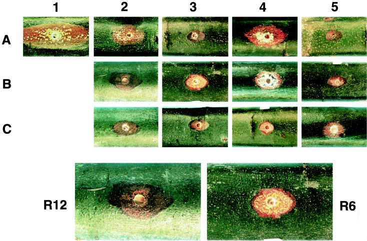
FIG. 4.
Representative cankers formed by virus-free and transfected C. parasitica isolates. The same coordinate system for strains transfected with chimeric viruses used in Fig. 2 is repeated here: EP155, A1; CHV1-Euro7, A2; CHV1-EP713, A3; R13, A4; R14, A5; R12, B2; R6, B3; R10, B4; R5, B5; R7, C2; R3, C3; R8, C4; and R9, C5. Cankers were photographed 30 days postinoculation. Cankers caused by R12 and R6 transfected isolates are enlarged at the bottom of the figure to illustrate contrast and to allow a closer inspection of the stromal pustules that contain spore-forming bodies, termed pycnidia, and the ridged margins of the canker formed by the R6 transfectant.

