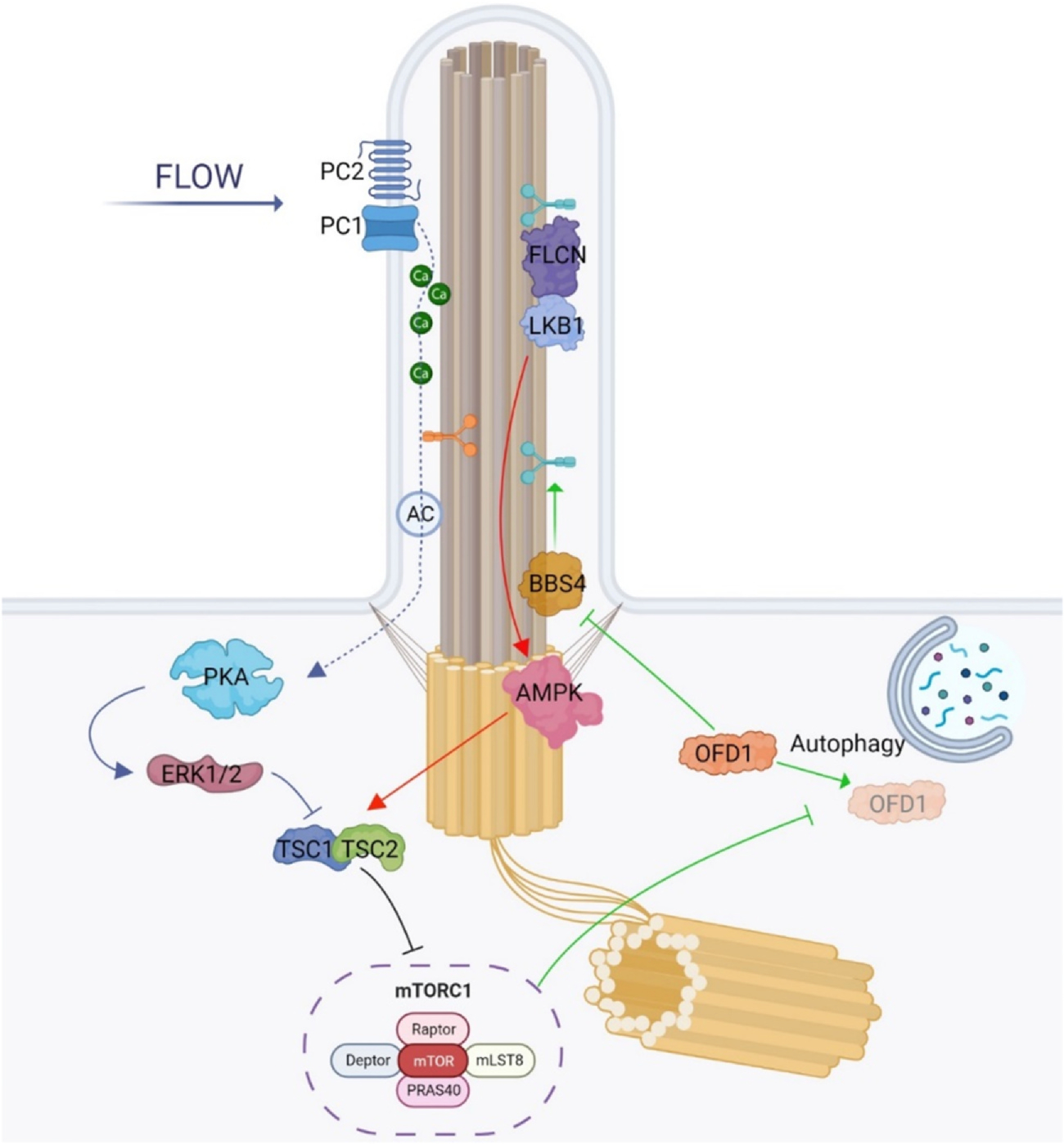
Fig. 2.

mTORC1 pathway in primary cilia: GREEN: Autophagy plays a role in regulating ciliogenesis. When there is a lack of nutrients and starvation conditions prevail, there is an increase in autophagy. This increase in autophagy promotes ciliogenesis by selectively degrading oral-facial-digital syndrome type 1 (OFD1), which is an inhibitor of BBSome formation. However, the autophagy-mediated degradation of OFD1 is negatively regulated by mechanistic target of Rapamycin complex 1 (mTORC1). The other two main mTOR/cilia related pathways rely on the flow stress-induced regulation of mechanistic target of Rapamycin complex 1 (mTORC1) as depicted in RED: Under flow stress conditions that cause primary cilia to deflect, mTORC1 activity reduces. This reduction is brought about by the accumulation of liver kinase B1 (LKB1) at the cilium through FLCN-mediated mechanisms. The accumulated LKB1 triggers the activation of AMP-activated protein kinase (AMPK) at the basal body, which subsequently phosphorylates TSC2, leading to the downregulation of mTORC1. BLUE: Polycystin-1 (PC-1) plays an important role in regulating mechanistic target of Rapamycin complex 1 (mTORC1). One of the main functions attributed to PC-1 is as a mechanosensor in primary cilia. When cilia are subjected to flow stress which results in bending, PC-1 is activated, which subsequently stimulates the ion channel PC-2, leading to calcium influx into the ciliary compartment. The ciliary calcium level increase in turn inhibits adenylyl cyclases (AC) and reduces the cyclic adenosine monophosphate (cAMP) level within the ciliary compartment. Conversely, when the flow stress is removed or there is dysfunction in PC-1, the calcium entry into the cilia is blocked, leading to a decrease in the ciliary calcium level. Thus, adenylyl cyclase activity increases, driving the ciliary cAMP level higher, which in turn activates PKA leading to the downregulation of mTORC1 through the ERK1/2 and TSC2.
