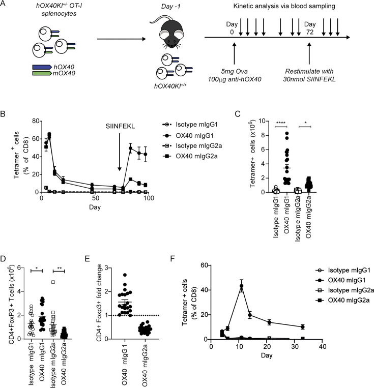
Figure 2.
anti-hOX40 mIgG1 and mIgG2a mAb are costimulatory in the OT-I/hOX40KI model. (A) Schematic of the experimental model; hOX40het OT-I cells are transferred into hOX40KIhom mice, immunized with ovalbumin in the presence of anti-hOX40 mAb and various cells measured by flow cytometry before recall stimulation with SIINFEKL. (B) Expansion of tetramer-positive OT-I cells in hOX40KIhom recipients. Isotype mIgG1, anti-hOX40 mIgG1 and anti-hOX40 mIgG2a n=4, Isotype mIgG2a n=3, representative of two independent experiments. Numeration of OT-I (C) and Treg (D) cells in hOX40KIhom spleens harvested on day 4 pooled from six independent experiments. Isotype mIgG1 and anti-hOX40 mIgG1 n=21, isotype mIgG2a and anti-hOX40 mIgG2a n=20, two mice per group were excluded due to no OT-I response. (E) Treg fold induction pooled from six independent experiments anti-hOX40 mIgG1 n=21 and anti-hOX40 mIgG2a n=20. (F) As in A except hOX40het OT-I purified CD8+T cells transferred in wildtype C57BL/6 recipients, n=4, representative of two independent experiments. C–E analyzed as one-way analysis of variance, Sidak’s multiple comparison. ****p<0.0001, **p<0.01, *p<0.05 mean±SEM. KI, knock-in; mAb, monoclonal antibody; Treg, regulatory T cells.

