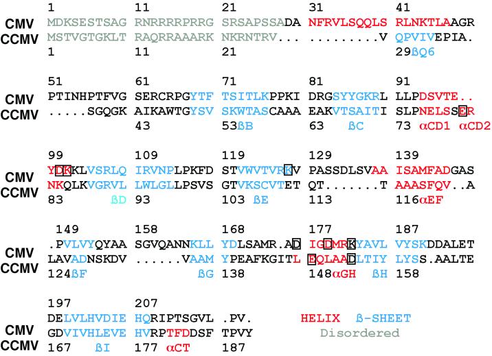
FIG. 3.
Sequence homology of CMV and CCMV based on structural alignments. The gray regions represent disordered regions, the red regions are helices, and the blue regions are β-strands. The nomenclature used for secondary elements is the same as that used for CCMV. The boxed amino acids are those involved in the subunit contacts about the quasi-threefold axes.

