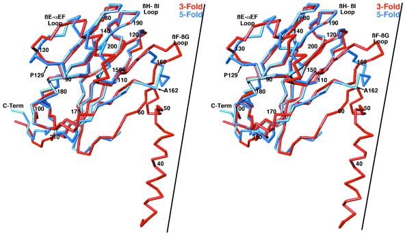
FIG. 5.
Comparison of the CMV A- and C-subunit structures. The C-α backbones of the A and C subunits are shown in blue and red, respectively. The RNA interior is toward the bottom of the diagram. The approximate locations of the threefold axis (for the C subunit) and the fivefold axis (for the A subunit) are represented by the black lines. C-Term, C terminus.

