FIG. 7.
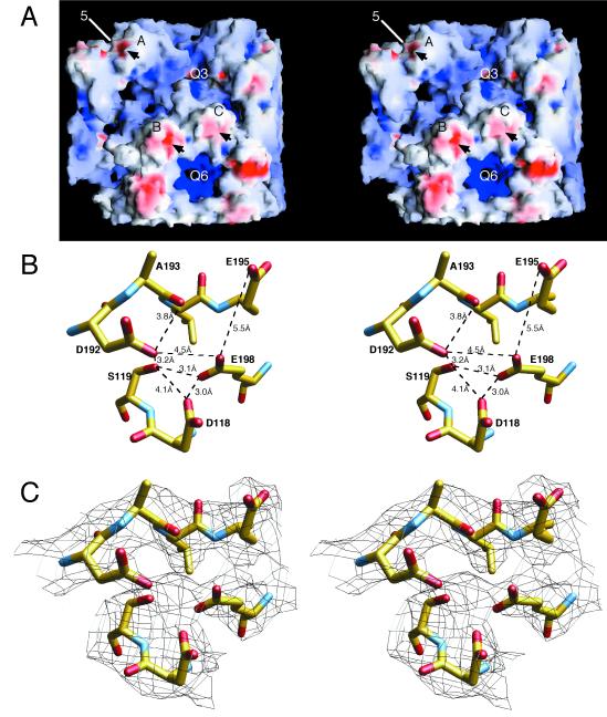
Structure of an external loop involved in aphid transmission. (A) van der Waals surface of a portion of the CMV capsid, with the negatively and positively charged electrostatic fields being shown in red and blue, respectively, created with the program GRASP (15). Arrows denote the locations of the loops described below. Note that the only negatively charged patch on the entire capsid surface is about the βH-βI loop. (B) Distances between the residues involved in this negatively charged patch. The atoms are colored according to atom type as defined in the legend to Fig. 5. The side chains in the area are very close to each other and may be indicative of a counterbalancing cation. (C) Same region and view as those shown in panel B, with the electron density contoured at 1 ς, represented by black lines. Note the patch of density between D118, S119, E198, and D192, which may represent a bound, divalent cation. The distances between the center of this patch of density and the oxygen atoms are between 2.3 and 3.0 Å. Upon deprotonation at neutral pH, these distances may decrease to those of typical oxygen-calcium contacts (∼2.3 Å). Q3 and Q6, quasi-three- and quasi-sixfold axes, respectively.

