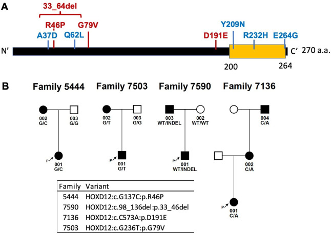
Figure 2.
Localisation and segregation of HOXD12 variants. (A) Localisation of HOXD12 variants identified in the study along the protein. HOXD12 variants co-segregated with clubfoot in multiplex families are marked in red. HOXD12 variants in singletons are marked in blue. Yellow motif represents homeobox domain. (B) HOXD12 variants co-segregate with clubfoot in four multiplex families with complete penetrance. a.a., amino acid.

