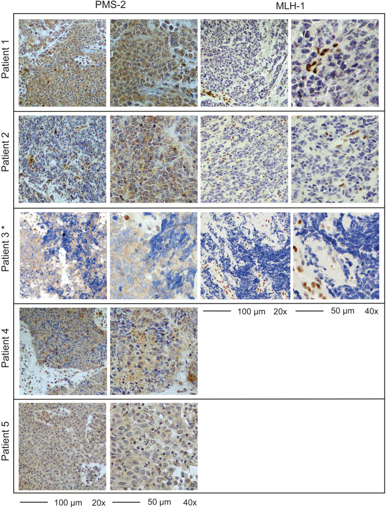
Figure 1.
Full section staining of PMS2 and MLH1 validates mismatch repair deficiency (MMR-D) in five patients. Representative images of full sections stained for MMR proteins with negative nuclear staining of tumor cells and positive staining of stroma or immune cells. Although some tumor cells exhibited cytoplasmic staining, no nuclear staining was found. This was confirmed by an expert pathologist. *Tissue block missing, photographs of tissue microarray (TMA).

