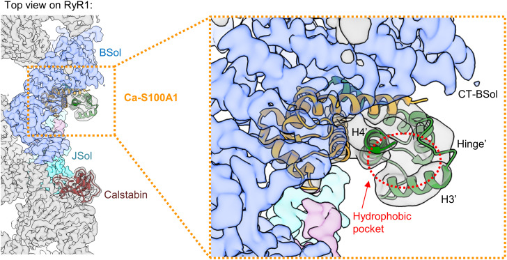
Fig. 3.
Hydrophobic pocket of the second monomeric subunit of S100A1 exposed at the cytosolic surface of RyR1. Top view on the cryo-EM composite map of RyR1 (gray) with Ca-S100A1 (subunit 1 in orange, subunit 2 in green; PDB: 8VK4) and Calstabin (brown). The S100A1 binding site of RyR1 is formed by JSol (cyan), BSol (blue), and SCL (magenta). In the magnification, the hydrophobic pocket of subunit 2 (hollow red circle), formed by the hinge region and helices H3′-H4′, is shown to be largely exposed at the surface next to the C-terminal BSol region (CT-BSol) while subunit 1 is bound to RyR1 beneath the BSol domain.

