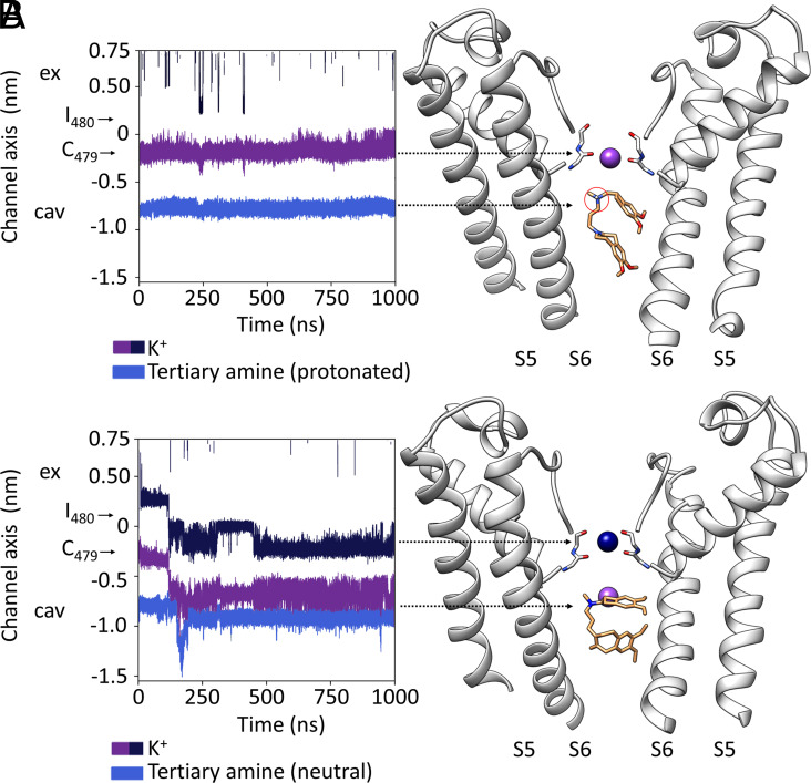
Fig. 5.
Mechanism of ivabradine block: (A and B) For simplicity, the present figure shows MD simulations of ivabradine pose 1, protonated (A) and neutral (B). MDs of pose 2 are, indeed, equivalent. (A, Left) Trajectory for a K+ ion (purple) in the SF and for the protonated tertiary amine of ivabradine (blue) in the pore cavity in a MD simulation performed with HCN4 pore only, in KCl solution. Positions of the carbonyl plane of C479 and I480 are indicated by black arrows. Ex (extracellular side of the pore); cav (intracellular pore cavity). (Right) Representative snapshot of the MD simulation shown in the Left panel, cross-referenced with the latter by arrowheads. For clarity, only two opposite subunits of the pore are shown. Over the entire simulation, the ion (purple) is stably located in the SF. Ivabradine (orange sticks) stably occupies the intracellular cavity. This localizes the protonated tertiary amine (circled in red) just below the SF for the entire simulation. (B, Left) Trajectories as in A, for K+ ions (purple and blue) in the SF and for the neutral tertiary amine of ivabradine (light blue). Positions of the carbonyl plane of C479 and I480 are indicated by black arrows. Ex (extracellular side of the pore); cav (intracellular pore cavity). (Right) Representative snapshot of the MD simulation shown in the left panel, cross-referenced with the latter by arrows. Entrance of a K+ ion (blue) from the extracellular side of the SF moves the second ion (purple) from the FS to the intracellular pore cavity. Neutralized ivabradine (orange sticks) stably occupies the intracellular cavity even when a K+ (purple) permeates the pore.

