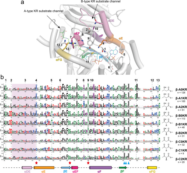
Figure 3.
(a) The position of fingerprint motifs in the KR structure (AmpKR2, A1-type, PDB ID 5XWV) [29]. Fingerprints are shown as sticks. The co-crystallized substrate mimic and co-factor are shown as lines. (b) Sequence logo comparation of the core moiety of β-module KRC based on the classification of their products. The key catalytic residues are marked by red stars, and the NADPH-binding residues (partially) are marked by blue triangles. The numbers at the top indicate the fingerprint motifs.

