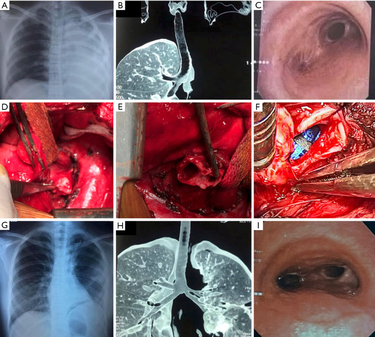
Figure 1.
Preoperative, intraoperative, and postoperative images of a case with left main bronchial stenosis. (A) Left lung collapse on preoperative chest X-ray; (B) left main bronchial occlusion on preoperative chest CT-scan with 3D reconstruction; (C) left main bronchial stenosis on preoperative bronchoscopy; (D) intraoperative image of left main bronchial occlusion; (E) anastomosis of the left main bronchus after resection of the stenotic segment; (F) anastomosis of the carina; (G) postoperative chest X-ray; (H) postoperative chest CT-scan with 3D reconstruction; (I) postoperative bronchoscopy image. CT, computed tomography.

