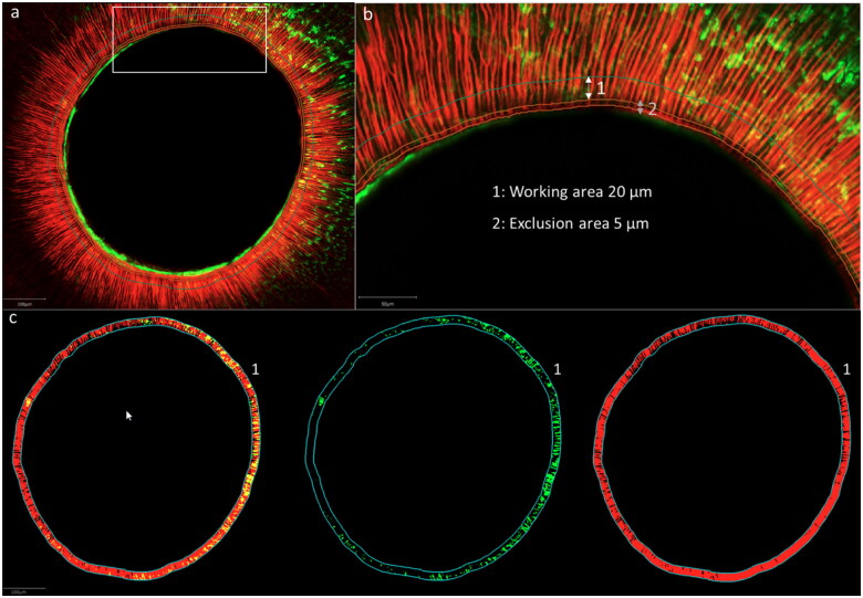
Figure 2.
Confocal images with an overview (a) of the root section, (b) magnification of the white box in (a) with 1: working area of 20μm and 2: exclusion of 5 μm; (c) working area with both fluorophores, fluorescein sodium salt (representing microleakage) and Rhodamine B isothiocyanate (representing infiltration) (from the left to the right)

