Figure 1.
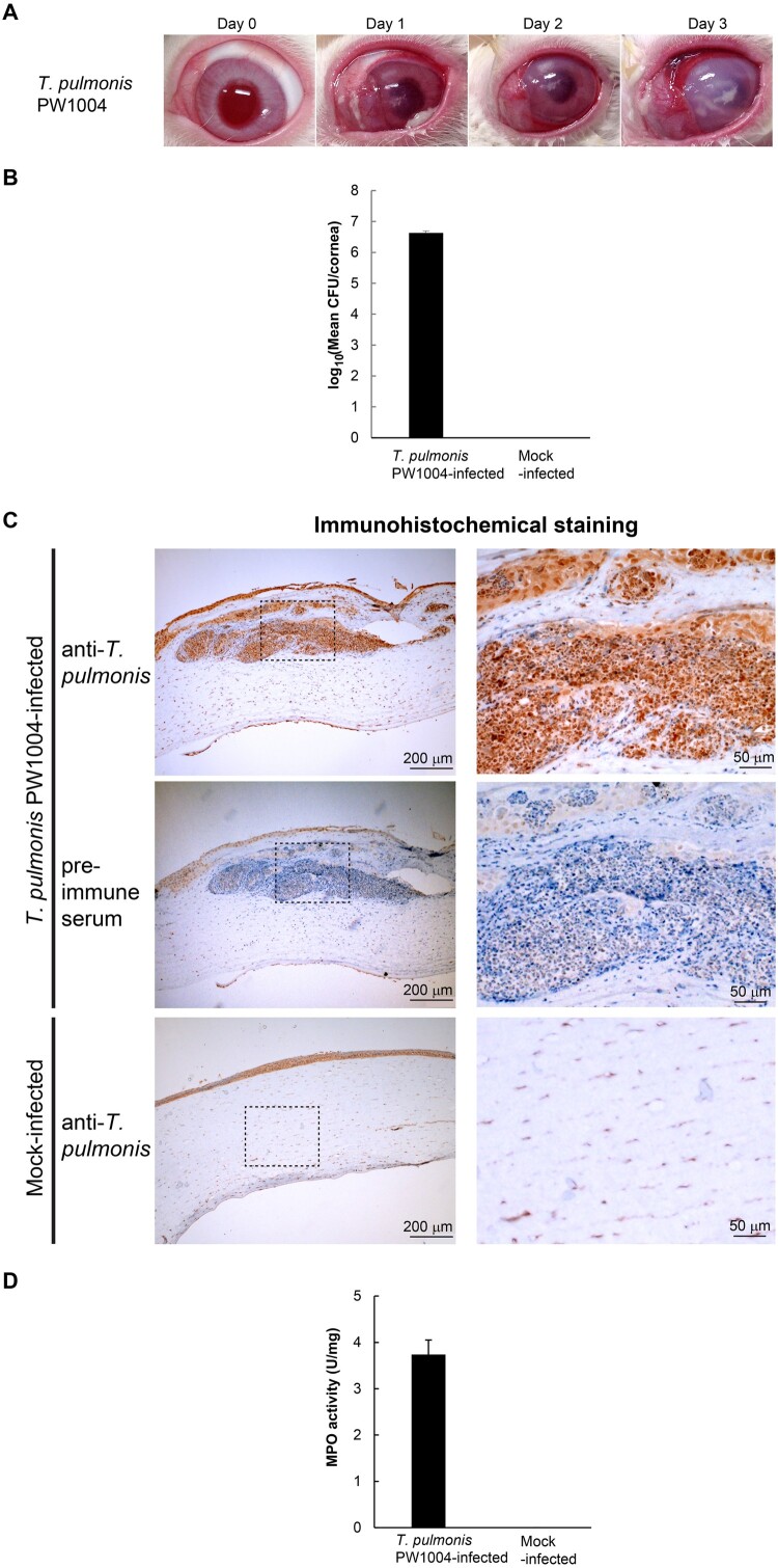
Experimentally induced keratitis in NZW rabbits after intrastromal injection of T. pulmonis-PW1004. (a) Gross appearance of the rabbit eyes after Tsukamurella infection. (b) Mean bacterial load recovered from the cornea of rabbits infected with T. pulmonis-PW1004 and those of control rabbits at 24 h PI. Error bars indicated mean CFU/cornea ± SEM of three independent experiments. (c) Immunohistochemical staining of corneal sections using mouse anti-T. pulmonis-PW1004 serum. The boxed area is further enlarged and shown in the right-hand panel of the corresponding image. Strong positive staining in brown colour against T. pulmonis could be detected in corneal sections from rabbits infected with T. pulmonis-PW1004 (top) but not from the mock-infected control rabbits (bottom). The middle panel shows corneal sections from the infected rabbits stained with pre-immune control serum; corneal sections of infected rabbits showing large amount of inflammatory cell infiltration with haematoxylin counterstain (top and middle). (d) MPO activity (U/mg) of the corneal tissues harvested from rabbits. Error bars indicate mean ± SEM of three independent experiments.

