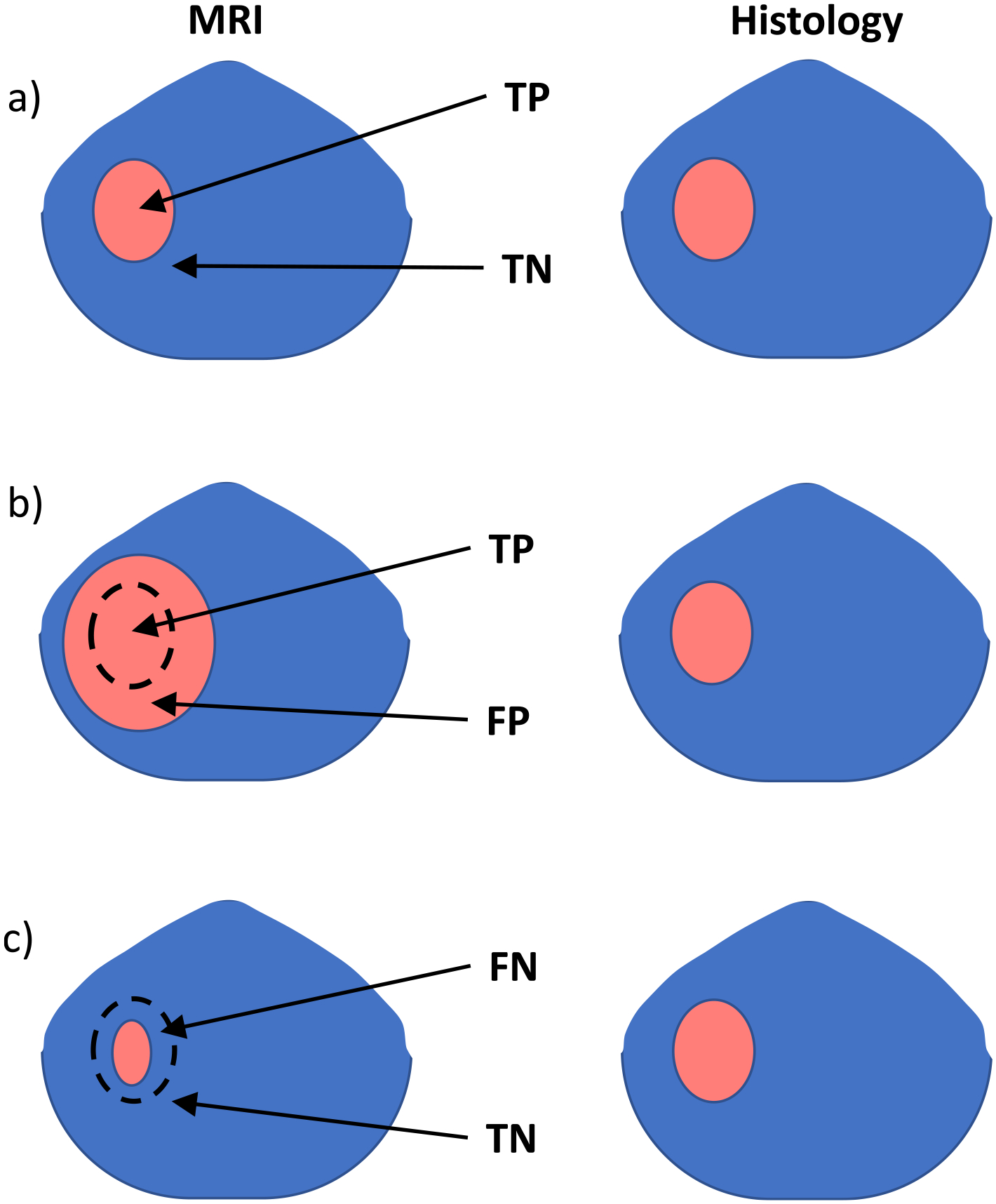
Fig. 2.

Examples of MRI lesion volume versus histology lesion volume with different classification results: a) TP when the MRI lesion size is within the outlier limits. b) FP when the MRI lesion size is larger than 1.33 • volumepathology + 1 cc. c) FN when the MRI lesion volume is <0.75 • (volumepathology − 1 cc).
