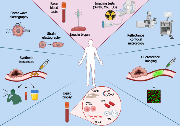
Figure 1.
Conventional and modern methods of cancer detection. The diagnostic path begins with learning the medical history of a patient, followed by performing basic blood tests (e.g., complete blood count, creatinine level, liver function test) and imaging tests (e.g., X-ray scan, ultrasound, magnetic resonance spectroscopy) to indicate where cancer may be located. Afterwards, the patient is scheduled for a needle biopsy. Prompt initiation of diagnosis is crucial since delayed cancer detection entails higher costs of treatment and hospitalization. Novel cancer detection methods include liquid biopsy, elastography, synthetic biosensors, fluorescence imaging, and reflectance confocal microscopy. Standards of cancer detection are located in a light pink area, whereas novel methods are placed on a light blue background. Figure created with BioRender. CTCs: Circulating tumor cells; CAFs: Cancer-related fibroblasts; TEPs: Tumor-educated platelets; cfDNA: Cell-free DNA; cfRNA: Cell-free RNA; EVs: Extracellular vesicles; US: Ultrasound (sonogram); MRI: Magnetic resonance imaging.

