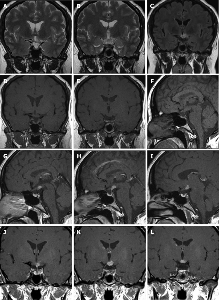
Figure 2.
Cranial magnetic resonance imaging enhancement scan showed pituitary height lower than normal for the same age group. Abnormal enhancement occurred anterior to the papillary body on the enhancement scan. A and B: Representative coronal T2-weighted images; C: Coronal fluid attenuated inversion recovery image; D: E: Representative coronal T1-weighted images; F: Sagittal T1-weighted image; G-I: Representative sagittal enhancement scans; J-L: Representative coronal enhancement scans.

