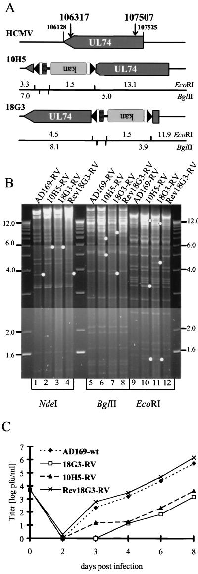
FIG. 7.
Mutant viruses defective in gene UL74 (gO) exhibit a reduction in growth kinetics. (A) Schematic representation of the Tn insertions. kan, ORF encoding the kanamycin resistance marker; upside down lettering indicates opposite orientation. Boldface numbers, exact nucleotide positions of the interruption of the ORF; lightface numbers, expected fragment sizes in kilobase pairs. (B) Viral DNA isolated from cells infected with AD169-RV (lanes 1, 5, and 9) compared to that from the UL74-deficient mutant viruses reconstituted from clones 10H5 (lanes 2, 6, and 10) and 18G3 (lanes 3, 7, and 11), together with that from a revertant genome generated by allelic exchange in bacteria and reconstituted to yield Rev18G3-RV (lanes 4, 8, and 12). Viral DNA was digested with NdeI (lanes 1 to 4). The UL74 gene is located on a 3.8-kbp NdeI fragment (lane 1), which, due to the Tn insertion in variants 10H5 and 18G3, shifts to a 5.6-kbp fragment (lanes 2 and 3, dots). In lanes 5 to 8 (BglII digests) and lanes 9 to 12 (EcoRI digests) the 18G3 revertant has a pattern indistinguishable from that of AD169-RV, whereas both gO-deficient viruses show the expected altered fragment sizes depicted in panel A (dots). (C) Single-step growth curve of gO mutants compared to revertant virus Rev18G3-RV. Confluent monolayers of MRC-5 cells were infected at an MOI of 0.01. Virus was harvested from both media and infected cells at the indicated time points and titered by plaque assay. Values shown are the results of duplicate assays from duplicate infections.

