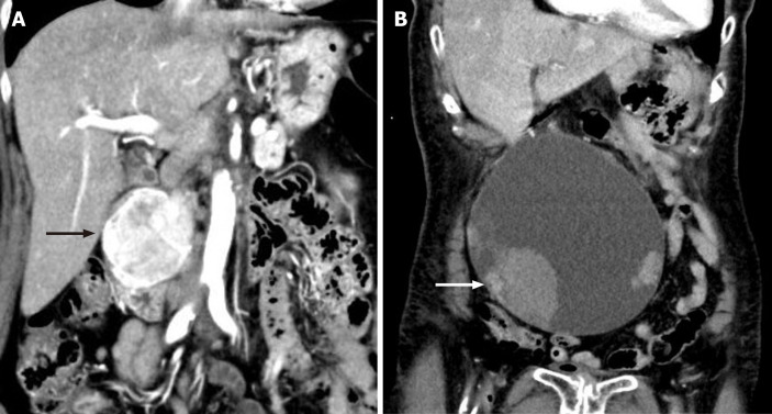
Figure 1.
Imaging of the duodenal tumor and pancreatic tumor. A: Imaging of the duodenal tumor and pancreatic tumor. Contrast-enhanced computed tomography showed a well-defined, enhancing masse, sized 5.0 cm × 6.0 cm, with heterogeneous density at the second portion of the duodenum (arrow); B: Imaging of the pancreatic tumor. Contrast-enhanced computed tomography showed a huge cystic mass measuring 17 cm, with some solid components and adjacent hematoma (arrow).

