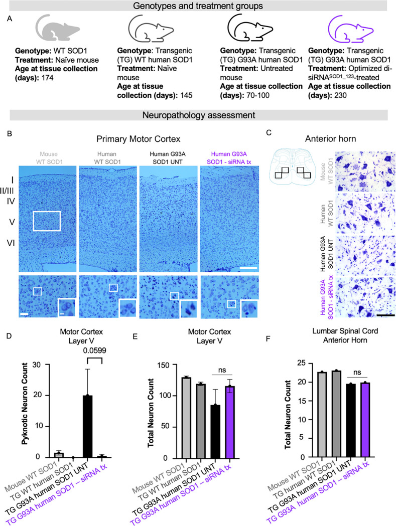
Figure 6. SOD1 knockdown with di-siRNA decreases number of apoptotic dark neurons in layer V of primary motor cortex in G93A mice.
(A) Genotypes and treatment groups used to assess neuropathology (B) Representative images of primary motor cortex, rectangles indicate the approximate region of interest (ROI) in layer V used for quantitative analyses. Scale bar = 250 μm (low power magnification), Scale bar=50 μm (high power magnification). (C) Cartoon and representative images showing ROIs used for quantification of pyramidal neurons. Scale bar=100 μm. (D) total number of Nissl-stained cortical neurons showed no statistical difference between groups. (E) dark neurons were significantly higher in number in the hSOD1G93A mice without treatment compared with other groups. (F) Number of pyramidal neurons counted within four ROIs in the anterior horn (cartoon) (average from two sections per animal). Data in the bar graphs are shown as mean ±SEM n=2 per group.

