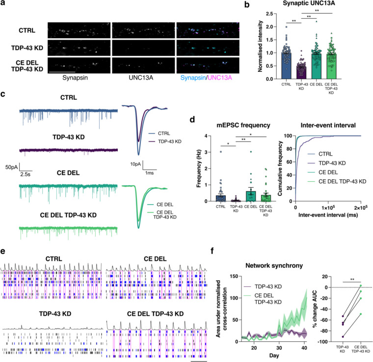
Fig. 3 |. Genomic deletion of UNC13A CE rescues synaptic function following TDP-43 knockdown.
a, Immunofluorescence labelling of synapsin and UNC13A pre-synaptic terminals in 4-week old HaloTDP and UNC13A CE Del iNeurons grown on rat astrocytes following 2-weeks of TDP-43 knockdown. Scale bar = 10 μm. b, Quantification of UNC13A intensity at synapsin positive synaptic terminals. Control n=71, TDP-43 knockdown n=71, CE Del n=66, CE Del TDP-43 knockdown n=75 synapses from 3 experiments. c, Representative mEPSC traces and average amplitude traces. d, Quantification of mEPSC frequency and inter-event interval cumulative distributions from control n=17, TDP-43 knockdown n=22, CE Del n=15, CE Del TDP-43 knockdown n=28 iNeurons pooled from 3 experiments. e, Representative raster plots from D39 multi-electrode array recordings from control, TDP-43 knockdown, CE Del, and CE Del TDP-43 knockdown conditions (30s window shown). Electrode bursts in blue and network bursts in pink. f, Time-course of network synchrony measured by the area under the normalised electrode cross correlation with TDP-43 knockdown conditions normalised to each control n=24 wells from 4 experiments. Graphs for (b) (d) (f) represent mean ± s.e.m. Statistics for (b) and (d) are one-way ANOVAs with Dunnet’s multiple comparisons test, statistics for (f) is a paired t test. *P < 0.05; ** P <0.01.

