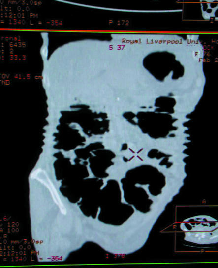
Figure 4.
Spiral computed tomography for colonography (this coronal reconstruction shows a sawtooth edge to the abdominal wall produced by breathing artefact). This study was performed on a spiral scanner. This artefact should be less common with multislice scanning.13 Computed tomographic colonography (virtual colonoscopy) was first introduced in the mid-1990s as a non-invasive technique to image the colon.14 Thin axial slices through the abdomen are obtained in supine and prone positions. These may be reconstructed into three dimensional (surface rendered) images giving the impression of viewing the large bowel via an endoscope. This technique will expand with further improvements in imaging technology and experience of observers.15

