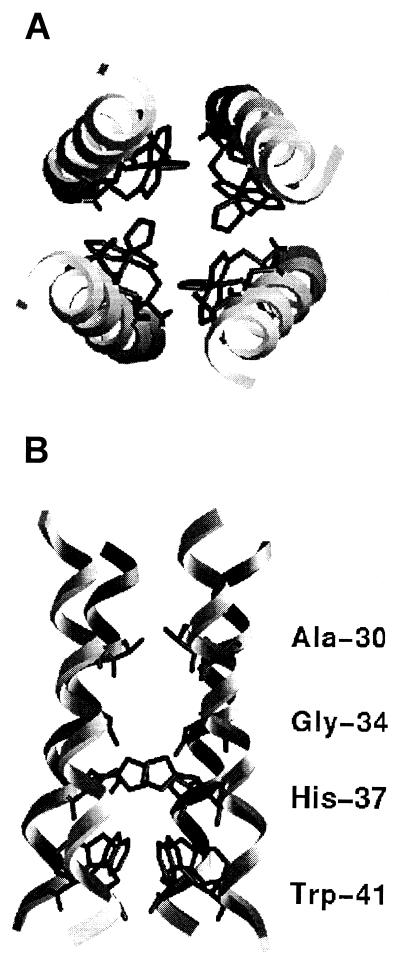
FIG. 5.
Model of the proposed TM domain of the M2 protein, showing top view as seen from the extracellular side (A) and a cross-section in the plane of the lipid bilayer (B). Residues which were identified as facing the ion-conducting aqueous pore are indicated. The model of the M2 channel is taken from that calculated in reference 20. The figure was generated using the program GRASP (18).

