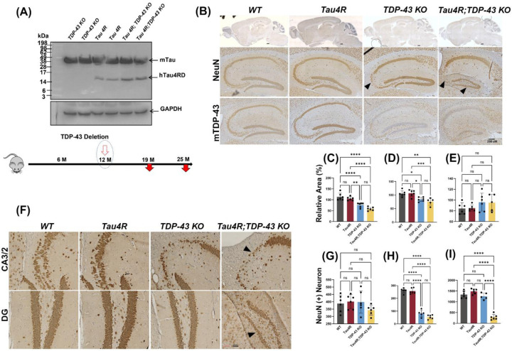
Figure 3. Loss of TDP-43 function accelerates neurodegeneration in hTau4R expressing mice.
(A) Immunoblot using a 77G7 antibody which recognizes the specific expression of hTau4RD fragments (~16kDa) including endogenous mouse tau protein expression. Bottom diagram shows the timeline for induction whereby TDP-43 is deleted at 12 months of age and mice are analyzed at either 19 or 25 months of age. (B & F) Immunohistochemical analysis of brains of 19-month-old WT (n=6), Tau4R (n=6), TDP-43 KO (n=5) and Tau4R;TDP-43 KO mice (n=6) using antiserum specific to NeuN to detect neurons; sagittal sections of whole brains (upper panel of B; scale bar, 500μm) and hippocampi (lower panels of B; scale bar, 200μm), F; magnified views of NeuN immunohistochemistry in hippocampal subregions CA2/3 & DG arrow heads indicates loss of neurons (both panels; scale bar, 20μm). Lower panel of B; immunohistochemistry of mouse TDP-43 showing depleted TDP-43 in mouse hippocampus (scale bar, 200μm). (C-E) Analysis of relative area measurements of hippocampus, cortex and cerebellum respectively depicted (% relative area of WT) (one-way ANOVA; ns: no significant difference; *P=0.0111 (WT vs TDP-43 KO) *P=0.0168 (Tau4R vs TDP-43 KO) **P=0.0014; ****P<0.0001). (G-I) Neuronal cell count of CA1, CA2/3 and DG regions from 19-month-old cohort (one-way ANOVA; ns: no significant difference; ****P<0.0001).

