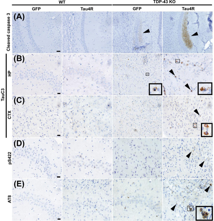
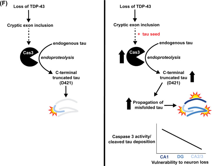
Figure 7. Caspase activation, tau cleavage and downstream tauopathy are further upregulated in mice lacking TDP-43 and expressing higher levels of hTauRD.
(A-E) Immunohistochemistry of brain sections of 18-month-old WT mice injected with GFP (n=3), WT mice injected with Tau4R (n=4), TDP-43 KO mice injected with GFP (n=4), and TDP-43 KO mice injected with Tau4R (n=4) mice using antiserum against (A) cleaved caspase 3 in the CA1 region of the hippocampus (scale bar, 50μm), (B-C) caspase-cleaved tau (TauC3) in the CA2/3 region of the hippocampus and cortex directly above hippocampus (scale bar, 20 μm), and (D) phosphorylated S422 and (E) AT8 in the CA2/3 region (scale bar, 20μm). (F) Model depicting that loss of TDP-43 splicing repression of cryptic exons precedes caspase 3-mediated cleavage of endogenous tau and eventual degeneration of CA2/3 neurons upon loss of TDP-43 in the mouse hippocampus. In the presence of a tau seed, elevated caspase 3-mediated cleavage of tau drives pathological tau aggregation and extends neuronal vulnerability to dentate granule neurons and CA1 neurons.


