Figure 1.
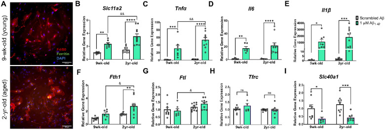
Age and Aβ stimulation synergize to increase microglial Slc11a2 and iron-loading markers in primary microglia.
A) Representative images of Percoll-isolated glia from young (top image, 9-week-old) and aged (bottom image, 2-year-old) mouse showing ferritin deposits in microglia from the aged mouse. Isolated glia were stained with antibodies raised against ferritin-L and F4/80, along with DAPI to visualize ferritin, microglia, and nuclei, respectively. Images shown at 20x, scale bar = 100 μm. B-I) Relative gene expression (compared to control scrambled Aβ) of B Slc11a2, C Tnfα, D Il6, E Il1β, F Fth1, G Ftl, H Tfrc, and I Slc40a1 via RT-qPCR. Isolated cells from young and aged mice were plated and treated with scrambled Aβ or 1μM Aβ1–42 for 24 h before collection for RNA isolation and RT-qPCR analysis. Two-way ANOVA, *p<0.05, **p<0.01, ***p<0.001, ****p<0.0001 effect of treatment. &p<0.05, &&p<0.01 effect of age × treatment. ns = not significant. Data represent the mean ± S.E.M of 7–11 mice per group. Statistical outliers were removed using the Grubb’s test.

