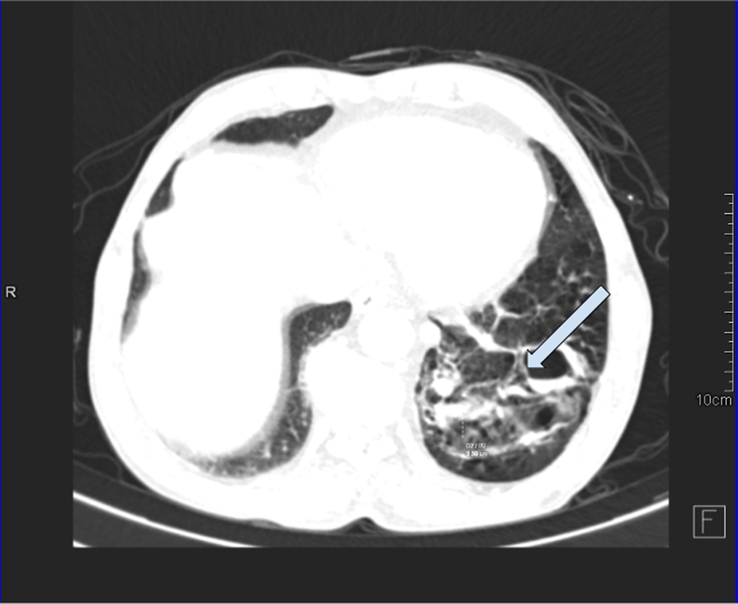
Figure 2.

Axial CT scan imaging in the lung algorithm showing multicystic lesion in the left lower lung lobe with a few intralesional cysts showing an air-fluid level and supplied by an aberrant artery arising from the descending thoracic aorta-pulmonary sequestration (infected) with cystic degeneration. COPD (chronic obstructive pulmonary disease) changes with centrilobular emphysema and cardiomegaly were noted in bilateral lung fields.
