Figure 3.

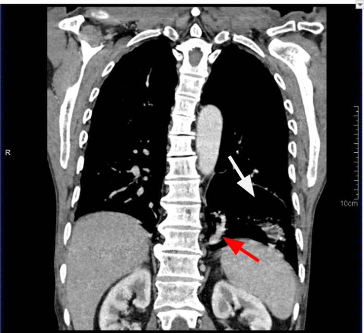
Chest coronal section contrast-enhanced computed tomography in mediastinal algorithm showing a section of the pulmonary sequestration as a cystic lesion (white arrow) with the systemic feeder aberrant artery from the aorta (red arrow).

Chest coronal section contrast-enhanced computed tomography in mediastinal algorithm showing a section of the pulmonary sequestration as a cystic lesion (white arrow) with the systemic feeder aberrant artery from the aorta (red arrow).