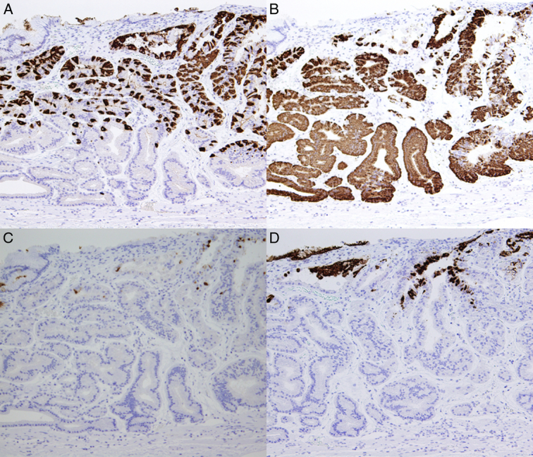
Figure 2.

Immunohistochemistry specimen for gastric carcinoma of the fundic gland type. (A) Pathological examination of the specimen showed that pepsinogen 1 was localized in the mucosa. (B) Pathological examination of the resection specimen showed that MUC6 was confined to the mucosa. (C) Tumor cells were positive for H+/K+-ATPase immunostaining. (D) Tumor cells were negative for MUC5AC.
