FIGURE 3.

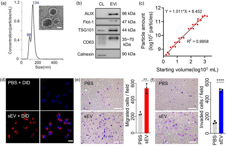
Characterization of EVi. (a) Size distribution (using NTA) and TEM image (inset) of MDA‐MB231 cell‐derived sEVs prepared using EVi system. Scale bar, 50 nm. (b) Immunoblot of various proteins in sEVs and whole‐cell lysates (CL) from MDA‐MB231 cells. One µg of protein was loaded in each lane. (c) Characterization of the particle yield as a function of the sample processing volume (n = 3). (d) Uptake of DID‐labeled sEVs isolated using EVi was detected using a confocal microscope. Scale bar, 20 µm. (e, f) Effect of MDA‐MB‐231 cell‐derived sEVs on MCF7 cells. Cell migration (e) and invasion assays (f) were performed (n = 3). Scale bar, 200 µm. Data are presented as means ± SD. *P < 0.05, ** P < 0.01, *** P < 0.001, and **** P < 0.0001, respectively; ns, not significant; unpaired two‐sided t‐test.
