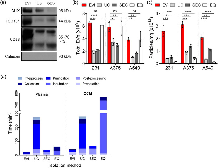
FIGURE 4.

Comparison of EVi with other isolation methods. (a) Quantitative immunoblot of sEV markers for MDA‐MB231 cell‐derived sEVs obtained using EVi, UC and SEC. 4 µg of proteins were loaded in each lane. (b, c) Total yields (b) and purities (c) were characterized using NTA analysis and BCA assay for each isolation method in three different cancer cell lines (n = 3), as indicated. 231, MDA‐MB‐231 breast cancer cell line; A375, A375 melanoma cell line; and A549, A549 lung cancer cell line. An equal volume of CCM (30 mL) was used to isolate sEVs according to each isolation method. (d) Comparison of the sample processing time by EVi and other isolation methods. Processed volumes were 500 µL for plasma and 200 mL for CCM, respectively. CCM, clarified conditioned media. Data are presented as means ± SD. *P < 0.05, ** P < 0.01, *** P < 0.001, and **** P < 0.0001, respectively; ns, not significant; unpaired two‐sided t‐test.
