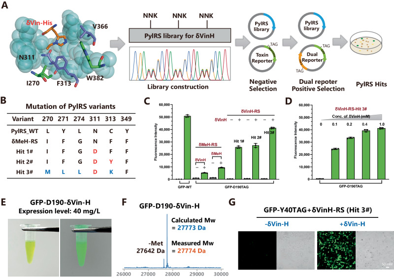
Fig. 3. Evolution of PylRS for the genetic encoding of δVin-H.
A The complex structure of δMeH-RS and AMP-δVin-H. The structure was regenerated from the pdb file 3qtc. B Evolved δVin-H-RS mutant for the recognition of δVin-H. C, D Validation of the selected variants through amber codon suppression of GFP-D190TAG in DH10B E. coli. The data in C, D are presented as mean values ± SD (n = 3, independent experiments). E Image of purified GFP-D190-δVin-H; the expression level reached 40 mg/L. F Validation of the δVin-H-incorporated GFP via LC‒MS. G Images of the incorporation of δVin-H into GFP-Y40TAG in HEK 293 T cells. Source data are provided as a Source Data file.

