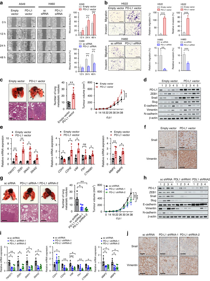
Fig. 4. PD-L1 overexpression enhances cell migration and invasion in vitro and promotes tumor progression and metastasis in vivo independently of anti-tumor immunity.
A549 and H522 cells were transfected with PD-L1-expressing or empty vector, and H460 and H596 cells were transfected with PD-L1 siRNA or scrambled siRNA. Then the cells were submitted to (a) a wound healing assay, and (b) cell migration invasion assay using Transwells. The wound closure rate was calculated using ImageJ. Scale bar = 1000 μm. The traversed cells after 24 h incubation were counted using ImageJ. Scale bar = 1000 μm. Histograms represent values normalized to control. PD-L1-overexpressing or vector control LLC cells and PD-L1-knockdown or shRNA control LLC cells were injected into the mammary fat pads of Balb/c-nu mice. c, g The primary tumor volume was measured with calipers every 2 or 3 days and calculated using the standard formula. Lung metastases were dissected 38 days after cancer cell injection, and tumor lung nodules (arrows) were counted. Representative hematoxylin- and eosin-stained images of lung tissues showed metastatic lesions. d, e, h, i mRNA and protein expression of EMT markers in lung tissues were analyzed by qRT-PCR and Western blotting. Numbers in (d) and (h) denotes individual mouse. f, j Representative IHC staining images for Snail and vimentin of lung tumors. Data in histograms are presented as mean ± S.E.M. of three independent experiments. *p < 0.05, **p < 0.01, and ***p < 0.001.

