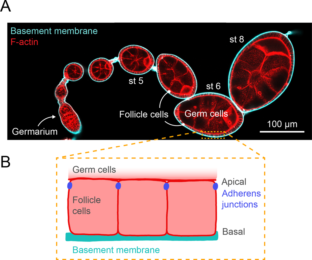
Fig. 1. Drosophila egg chamber organization.
A. Confocal cross-section image through an ovariole stained for F-actin (phalloidin) and expressing GFP-Collagen IV to label the basement membrane. Note that different sizes of egg chambers require different levels of compression to hold them in place in the flow chamber. B. Illustration of the organization of the follicular epithelial cells that surround the egg chamber. The apical surfaces of follicle cells contact the germline cells and the basal surfaces contact the basement membrane on the exterior of the tissue.

