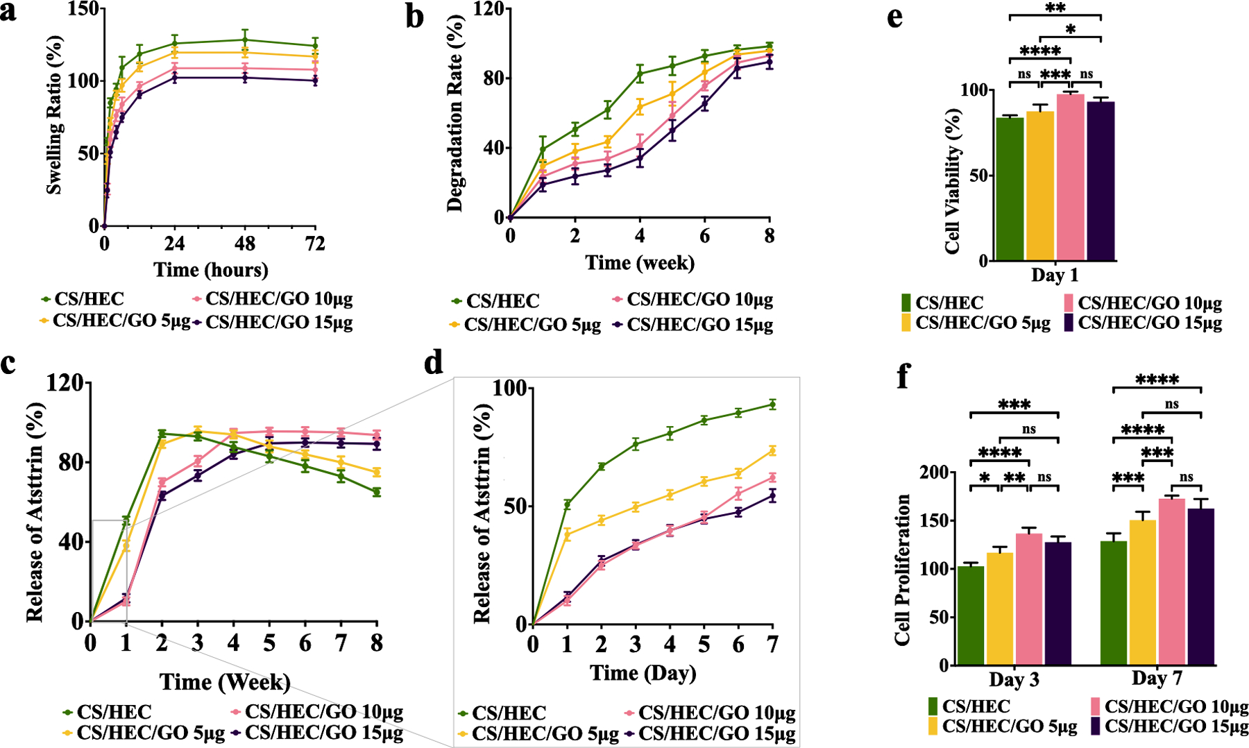
Fig. 3. In vitro characterization of hydrogels.

a) The swelling rate of the prepared CS/HEC, CS/HEC/GO 5 μg, CS/HEC/GO 10 μg, and CS/HEC/GO 15 μg hydrogels in PBS within 72 hours. b) The degradation rate of the prepared CS/HEC, CS/HEC/GO 5 μg, CS/HEC/GO 10 μg, and CS/HEC/GO 15 μg hydrogels in PBS within 8 weeks. c) The release curves represent Atsttrin release into PBS at 37 °C over 8 weeks from CS/HEC, CS/HEC/GO 5 μg, CS/HEC/GO 10 μg and CS/HEC/GO 15 μg hydrogels. d) Represents the data for the first 7 days or the first week. The experiments were done in 3 replicates. e) The cytocompatibility evaluation of the hydrogels in contact with bmMSCs by MTT assay at 24 hours post-incubation. f) Cell proliferation assay at day 3 and 7 after incubation. Two-way ANOVA shows significant differences between the experimental and control group (*p < 0.05; **p < 0.01; ***p < 0.001; ****p < 0.0001). The experiments were done in 5 biological replicates (n=5), and the data presented are mean ± SD.
