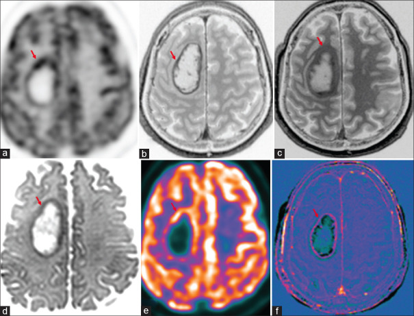
Figure 1.
Case of cerebral metastases in primary lung malignancy shows increased marginal uptake in right frontal lobe lesion (red arrow) in 18F-fluorodeoxyglucose(FDG) PET-AC(attenuation corrected) (a) and color-coded positron emission tomography images (e), peripheral enhancement (b) and washout in 75-min delayed postcontrast (c), marginal diffusion restriction (d) and marginal enhancement in subtracted T1 contrast image (f)

